

[ps2id id=’background’ target=”/]
BACKGROUND
Western blotting is an essential technique to analyze protein in various studies such as cellular and molecular biology and biochemistry in biomedical research. Post-translational modification, protein phosphorylation and protein synthesis can be investigated with the help of this technique.
The aim of experiment is to execute western blot successfully from the animal cells.
[ps2id id=’requirements’ target=”/]
REQUIREMENTS
Sample: Protein sample (Animal cells).
Apparatus: Cell scraper,
Eppendorf tubes,
Spectrophotometer,
Biorad gel electrophoresis system,
Rack set for the membrane transfer system.
Chemicals: NP-40,
NaCl,
Tris-HCl,
Triton X100,
Sodium deoxycholate,
Sodium dodecyl sulphate (SDS),
Ammonium persulfate,
2-Mercaptoethanol,
Glycerol,
Bromophenol blue,
Glycine,
Methanol,
BSA (bovine serum albumin),
TEMED,
Polyvinylidene fluoride (PVDF) membrane,
Primary antibody,
Secondary antibody (Horse radish peroxidase, HRP),
1X PBS,
1X TBST.
Required Buffers3
There are several steps for the completion of this technique where many buffer were used to perform. They are as follows:
-
NP-40 buffer
150 mM NaCl
1.0% NP-40 (possible to substitute with 0.1% Triton X-100)
50 mM Tris-HCl, pH 8.0
Protease inhibitors
-
RIPA buffer (radioimmunoprecipitation assay buffer)
150 mM NaCl
1.0% NP-40 or 0.1% Triton X-100
0.5% sodium deoxycholate
0.1% SDS (sodium dodecyl sulphate)
50 mM Tris-HCl, pH 8.0
Protease inhibitors
-
Tris-HCl
20 mM Tris-HCl
Protease inhibitors
-
Laemmli 2X buffer/loading buffer
4% SDS
10% 2-mercaptoethanol
20% glycerol
0.004% bromophenol blue
0.125 M Tris-HCl
Check the pH and adjust to 6.8
-
Running buffer (Tris-Glycine/SDS)
25 mM Tris base
190 mM glycine
0.1% SDS
Check the pH and adjust to 8.3
-
Transfer buffer (wet)
25 mM Tris base
190 mM glycine
20% methanol
Check the pH and adjust to 8.3
For proteins larger than 80 kDa, final concentration of SDS 0.1%.
7–
Blocking buffer
3–5% milk or BSA (bovine serum albumin)
Add to TBST buffer. Mix well and filter. Failure to filter can lead to spotting, where tiny dark grains will contaminate the blot during color development.
Composition of casting gel for gel electrophoresis
| Components (10% resolving gel for SDS-PAGE) | Volume of components required for the gel (ml) total volume 20 ml |
| H2O 30% acrylamide mix 1.5M Tris (pH8.8) 10% SDS 10% ammonium sulfate TEMED |
7.9 6.7 5.0 0.2 0.2 0.008 |
[ps2id id=’procedure’ target=”/]
PROCEDURE
Cell lysate preparation from Animal cells (all the process should be executed on the ice)
Get the culture dish from incubator and place on ice, wash with confluent cells with PBS (ice-cold). Discard the PBS, add the lysis buffer (stored in 4°C) in the cell culture petri dish (600µl/60mm culture dish). Scrape all the attached cells from the dish with cell scraper, further safely collect the cell suspension into an Eppendorf tube. Constantly vertex for 30 min or more at 4°C and centrifuge at 12000rpm for 20-30 min (It can vary from cell to cell). Keep the tube on ice, transfer the supernatant in a fresh tube and discard the pellet.
Take a small fraction of lysate to quantify the concentration of protein in spectrophotometer, protein concentration for each sample lysate from animal cells should be quantified and calculated before adding the two fold of Laemmli buffer. Load similar volume of proteins with buffer. Boil the samples for 5 min at 100°C to denature in the sample buffer further aliquot the lysates and stored at -20°C for the future applications.
Casing gel
The gel, which we are casting, is for 15-100kDa proteins. The gel casting set is from Biorad. Following is the gel composition.
Loading and running the gel (SDS-PAGE) electrophoresis
Equal concentration of protein should be loaded in wells of the SDS-PAGE gel, based on molecular weight marker. Total protein of 20–30 μg from cell lysate further gel running for 1–2 h at 100 V. Optimization of voltage and time for gel running is needed.
Transferring the protein on the membrane
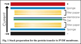 PVDF membrane should be activated with methanol for 1-3 min and rinse with transfer buffer prior to stalk preparation. Ponceau S staining is a method for inquiring protein on the membrane prior to the blocking step.
Execute membrane blocking with TBST buffer containing 5% BSA, at room temperature for 1 h, further membrane incubation in appropriate dilutions (1:40,000) of primary antibody in blocking solution for overnight at 4°C. Membrane washing with TBST buffer, three times per 5 min. Membrane incubation with the dilution (Recommendation based on secondary antibody) of conjugated secondary antibody in blocking solution at RT for 1 h. Wash the membrane with TBST for 5 min each three washes. Drain the excess reagent attached on membrane and further cover the membrane in transparent plastic wrap. Chemiluminescence colorimetric detection should be developed in darkroom.1-3
[ps2id id=’conclusion’ target=”/]
CONCLUSION
This experiment concluded the western blotting technique in for the cells from animal cells.
[ps2id id=’references’ target=”/][ps2id id=’1′ target=”/]
REFERENCES
-
Kumar A, Mishra HK., Dwivedi P., Subramaniam. Secreted trophic factors of Human umbilical cord stromal cells induce differentiation and neurite extension through PI3K and independent of cAMP pathway. Annals of Neuroscience 2015;22(2):97-106.
-
Tahrin Mahmood and Ping-Chang Yang. Western Blot: Technique, Theory, and Trouble Shooting. North American J Medical Sciences. 2012;4(9):429-34.